


Download the article in pdf format
Publication date: 01.06.2023
DOI: 10.24412/2782-6570-2023_02_02_3
UDC 796.015.527
INFLUENCE MECHANISMS OF TRAINING WITH BLOOD FLOW RESTRICTION ON HYPERTROPHY OF WORKING MUSCLES: A SCOPING REVIEW
F.A. Koloskov, A.B. Miroshnikov, A.V. Meshtel’
Russian State University of Physical Education, Sport, Youth and Tourism, Moscow, Russia
Annotation. Although the hypertrophic effects of resistance training and blood flow restriction have been demonstrated by numerous studies, the underlying mechanisms responsible for these effects have not been elucidated. Objective: to conduct a scoping review of the available literature to analyze and summarize the mechanisms underlying muscle hypertrophy during training with blood flow restriction. The systematic search of publications in PubMed ended on 05.05.2023. This study was conducted in accordance with the statement of Preferred Reporting Items for Systematic reviews and Meta-Analyses extension for Scoping Reviews (PRISMA-ScR). A total of 128 references were identified. As a result of selection, 6 reviews were analyzed and included in the study. Systematizing the data, we came to the conclusion that the increased level of metabolic stress is the main driving mechanism of muscle growth during blood flow restriction training.
Keywords: occlusion training, blood flow restriction training, muscle hypertrophy.
Introduction. Blood flow restriction (BFR) training was developed in Japan in the late 70s, where it was called the KAATSU training [1]. Although long-term benefits of the BFR exercises, including increase of muscle hypertrophy, strength and improved aerobic and anaerobic working capacity are clearly described in scientific literature, a discussion on underlying mechanisms responsible for adaptation is still going on.
In terms of muscle hypertrophy, the occurring hypertrophic effects of strength training with BFR are due to an increased level of metabolic stress (i.e. metabolite accumulation as a response to ischemic/hypoxic environment) [2], which in theory causes muscle growth, influencing other factors, such as HTMU's (high threshold motor units) recruitment [3-4], increased level of systemic hormones [5-6], cell edema [7] and increased production of ROS (reactive oxygen species) [8].
Since many researchers explained possible mechanisms underlying muscle growth after BFR training in different ways, and the data cannot be merged into either a systematic or umbrella review, we have set an objective of our study.
The objective: to conduct a scoping review of the available literature to analyze and summarize the mechanisms underlying muscle hypertrophy during training with blood flow restriction.
Methods and organization. The study took place in the Department of Sports Medicine (Russian State University of Physical Education, Sport, Youth and Tourism, Moscow). It was conducted in accordance with PRISMA-ScR requirements (Preferred Reporting Items for Systematic reviews and Meta-Analyses extension for Scoping Reviews) [9]. The study’s protocol was made before the search and did not change either during or after the research. Prior to the search, we have determined that the scoping review would only include reviews of the subject field that examine mechanisms underlying muscle hypertrophy during BFR training.
The literature search was made in the PubMed database. The following keywords are: “blood flow restriction training AND (muscle mass OR muscle strength OR skeletal muscle hypertrophy OR muscle protein synthesis OR muscle growth)”; “KAATSU AND (muscle mass OR muscle strength OR skeletal muscle hypertrophy OR muscle protein synthesis OR muscle growth)”; “occlusion training AND (muscle mass OR muscle strength OR skeletal muscle hypertrophy OR muscle protein synthesis OR muscle growth)”. No time frames were set, but the last search was dated 05.05.2023.
In order for the research to be included, it must correspond with the inclusion criteria based on the PICOS system [10]. P (Population) – active and non-active participants, athletes, elite athletes (men and women) above 18; I (Intervention) – BFR training (strength training, training with weights); C (Comparison) – comparison with the control group or when the intervention starts; O (Outcomes) – mechanisms of BFR training’s influence on hypertrophy of working muscles were studied; S (Study) – reviews of the subject field, systematic reviews and meta-analyses, in which the aforementioned mechanisms were studied.
Initially, two authors of the review (F.A. Koloskov, A.B. Miroshnikov) simultaneously and independently checked titles of the articles, their abstracts and, when needed, full texts in accordance with the criteria. Duplicates and articles that did not correspond were excluded. There was no language barrier. Since all the data were presented in a descriptive way, there was no statistical analysis.
Results and discussion. A total of 128 mentions were found. Then we exported the needed data; all duplicates and irrelevant articles were deleted manually. The figure below shows the PRISMA scheme describing the selection process. Only 6 reviews corresponded with the criteria [11-16].
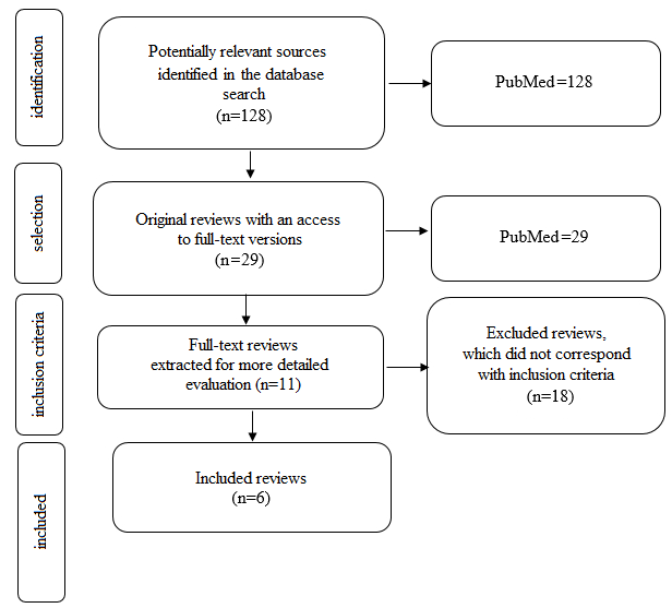
Fig. The PRISMA scheme
Hwang et al [11] identify following mechanisms contributing to muscle growth during BFR training:
1) metabolic accumulations, which can stimulate anabolic growth factors;
2) increased muscle protein synthesis through specific intracellular signaling pathways;
3) muscle injury;
4) mechanotransduction;
5) HTMU's recruitment patterns;
6) increased activity of satellite cells;
7) ROS production;
6) increased anabolic hormones’ (testosterone, growth hormone, insulin-like growth factor 1 – IGF1)) levels.
Moreover, the authors assume that mechanical load and metabolic stress can act synergistically to create suitable conditions for the greatest hypertrophic potential.
Loenneke et al [12] reveal 4 phases, in which BFR training is applied:
1) bed rest;
2) low-intensity walking;
3) low-intensity training with weights;
4) low-intensity exercises combined with high-intensity strength training.
During the first phase, the predominant mechanisms participating in support of skeletal muscle mass with BFR activate muscle cell edema and enhancement of signal transfer through β2-adrenergic receptors when there is no physical load. The predominant mechanisms that are identified during the 2nd phase are rapid increase in volume of muscle cells and
increased cardiac output or blood volume. The predominant mechanisms in the following phases are:
1) HTMU's recruitment;
2) additional secretion of anabolic hormones.
Pearson et al [13] divide mechanisms, with the help of which BFR training stimulates muscle growth, into primary and secondary ones.
The main (primary) mechanisms are mechanical load and metabolic stress. The authors assume that the primary factors will effect a number of connected secondary mechanisms of the muscle growth induction, such as mechanotransduction, muscle injury caused by physical exercises, increased secretion of systemic and localized hormones, cell edema, increased ROS and nitrogen oxide production (which may also lead to production of heat shock proteins), as well as HTMU’s recruitment. The authors also note potential autocrine (i.e. protein synthesis stimulation due to increase of anabolic and/or decrease of catabolic signaling pathways) and paracrine (i.e. increased activation, proliferation and fusion of satellite cells) mechanisms participating in hypertrophy caused by BFR training with weights.
Yuan et al [14] identify the following main mechanisms, with which BFT training stimulates skeletal muscle growth:
1) HTMU's recruitment;
2) activation of the protein synthesis signaling pathway;
3) metabolic stress;
4) anabolic hormones’ secretion;
5) cell edema.
Freitas and his colleagues [15] name the following mechanisms:
1) metabolic stress;
2) HTMU's recruitment;
3) anabolic hormone secretion;
4) increased protein synthesis due to changes in biomolecular pathways, including the mammalian target of rapamycin complex 1 (mTORC1) and inhibition of such atrogenes as the Muscle RING Finger1 (MuRF1) and atrogin-1, as well as myostatin pathway inhibition.
Vopat et al claim that the suggested mechanisms are based on two main factors: metabolic and mechanical stress [16].
These factors act synergetistically, signaling about a number of secondary mechanisms including tissue hypoxia, metabolite accumulation and cell edema. At the end, it contributes autocrine and paracrine signaling pathways leading to protein synthesis, HTMU's recruitment, local and systemic synthesis of anabolic hormones and myogenic stem cells’ stimulation.
When integrating information about all mechanisms (table) mentioned in the works and included into the scoping review, we can conclude the following: 83.3% – metabolic stress, 33.3% – mechanical load, 33.3% – mechanotransduction, 33.3% – muscle injury caused by training, 50% – increase of the protein synthesis due to mTORC1 changes, 100% – HTMU's recruitment, 100% – increased anabolic hormones’ levels, 33.3% – ROS production, 66.7% – muscle cell edema, 50% – increased satellite cell activity and 16.7% – myostatin pathway inhibition.
Table
Integration of information about all mechanisms, with which blood restriction training stimulate skeletal muscle growth
|
Mechanism |
Reference |
|
metabolic stress |
[11,13,14,15,16] |
|
mechanical load |
[11,13] |
|
mechanotransduction |
[11,13] |
|
muscle injury caused by training |
[11,13] |
|
increase of the protein synthesis due to mTORC1 changes |
[11,13,14] |
|
HTMU's recruitment |
[11,12,13,14,15,16] |
|
increased anabolic hormones’ levels |
[11,12,13,14,15,16] |
|
ROS production |
[11,13] |
|
muscle cell edema |
[12,13,14,16] |
|
increased satellite cell activity |
[11,13,16] |
|
myostatin pathway inhibition |
[15] |
Note: HTMU’s – high threshold motor units; ROS – reactive oxygen speciesа; mTORC1 – mammalian target of rapamycin complex 1
Considering the fact that the increased systemic hormones’ level will not probably effect muscle growth [17-18], mechanical load and metabolic stress are apparently the main mechanisms of muscle growth. Moreover, metabolic accumulations that occur as a result of metabolic stress may provide the additional mechanism of HTMU’s recruitment [19], which contributes to a creation of mechanical tension in these fibers. Eventually, almost the whole muscle growth process may be due to mechanical stress with slight influence of metabolic stress and possibly even harmfuleffect of muscle injury [20].
Conclusion. Although exercises with weights and BFR are indeed popular and effective, the mechanisms underlying hypertrophic adaptation have not yet been identified. While creating a data system, we have concluded that an increased metabolic stress level is the main stimulus in this process, which, as supposed, activates the other mechanisms (e.g. systemic hormone production, increased HRMU’s recruitment), all of which are believed to mediate muscle growth due to autocrine and/or paracrine action. However, the extent to which these mechanisms are activated under metabolic stress is unknown. Additional randomized controlled trials are required to determine the degree and contribution of metabolic stress to the creation of mechanical stress in high threshold motor units.
Conflict of interest. The authors declare no clear or potential conflicts of interest associated with the publication.
Authors’ contribution. Concept, design of the study, writing – F.A. Koloskov; data gathering – F.A. Koloskov, A.B. Miroshnikov; editing – A.V. Meshtel’.
REFERENCES
- Slysz J. Stultz J., Burr J.F. The efficacy of blood flow restricted exercise: A systematic review & meta-analysis. J Sci Med Sport, 2016, vol 19, no. 8, pp. 669-675. DOI: 10.1016/j.jsams.2015.09.005.
- Loenneke J.P., Fahs C.A., Wilson J.M., Bemben M.G. Blood flow restriction: the metabolite/volume threshold theory. Med Hypotheses, 2011, vol. 77, no. 5, pp. 748-752. DOI: 10.1016/j.mehy.2011.029.
- Moritani T., Sherman W.M., Shibata M., Matsumoto T., Shinohara M. Oxygen availability and motor unit activity in humans. Eur J Appl Physiol Occup Physiol, 1992, vol. 64, no. 6, pp. 552-556. DOI: 10.1007/BF00843767.
- Yasuda T., Brechue W.F., Fujita T., Shirakawa J., Sato Y., Abe T. Muscle activation during low-intensity muscle contractions with restricted blood flow. J Sports Sci, 2009, vol. 27, no. 5, pp. 479-489. DOI: 10.1080/02640410802626567.
- Chen Y., Wang J., Li S., Li Y. Acute effects of low load resistance training with blood flow restriction on serum growth hormone, insulin-like growth factor-1, and testosterone in patients with mild to moderate unilateral knee osteoarthritis. Heliyon, 2022, vol. 14, no. 8, p. 10. DOI: 10.1016/heliyon.2022.e11051.
- Vilaça-Alves J., Magalhães P.S., Rosa C.V., V.M. Reis, Garrido N.D., Payan-Carreira R., Neto G.R., Costa P.B. Acute Hormonal Responses to Multi-Joint Resistance Exercises with Blood Flow Restriction. J Funct Morphol Kinesiol, 2022, vol. 8, no. 1, p. 3. DOI: 10.3390/jfmk8010003.
- Loenneke J.P., Fahs C.A., Rossow L.M., Abe T., Bemben M.G. The anabolic benefits of venous blood flow restriction training may be induced by muscle cell swelling. Med Hypotheses, 2012, vol. 78, no. 1, pp. 151-154. DOI: 10.1016/j.mehy.10.014.
- Preobrazenski N. Islam H., Gurd B.J. Molecular regulation of skeletal muscle mitochondrial biogenesis following blood flow-restricted aerobic exercise: a call to action. Eur J Appl Physiol, 2021, vol. 121, no. 7, pp. 1835-1847. DOI: 10.1007/s00421-021-04669-6.
- Tricco A.C., Lillie E., Zarin W., O'Brien K.K., Colquhoun H., Levac D., Moher D., Peters M.D.J., Horsley T., Weeks L. PRISMA Extension for Scoping Reviews (PRISMA-ScR): Checklist and Explanation. Ann Intern Med, 2018, vol. 169, no. 7, pp. 467-473. DOI: 10.7326/M18-0850.
- Samson D., Schoelles K.M. Chapter 2: medical tests guidance (2) developing the topic and structuring systematic reviews of medical tests: utility of PICOTS, analytic frameworks, decision trees, and other frameworks. J Gen Intern Med, 2012, vol. 27. DOI: 10.1007/s11606-012-2007-7.
- Hwang P.S., Willoughby D.S. Mechanisms Behind Blood Flow-Restricted Training and its Effect Toward Muscle Growth. J Strength Cond Res, 2019, vol. 33, pp. 167-179. DOI: 10.1519/JSC.000000
- Loenneke J.P., Abe T., Wilson J.M. Thiebaud R.S., Fahs C.A., Rossow L.M., Bemben M.G. Blood flow restriction: an evidence based progressive model (Review). Acta Physiol Hung, 2012, vol. 99, no. 3. pp. 235-250. DOI: 10.1556/APhysiol.99.2012.3.1.
- Pearson S.J., Hussain S.R. A review on the mechanisms of blood-flow restriction resistance training-induced muscle hypertrophy. Sports Med, 2015, vol. 45, no. 2, pp. 187-200. DOI: 10.1007/s40279-014-0264-9.
- Yuan J., Wu L., Xue Z., Xu G., Wu Y. Application and progress of blood flow restriction training in improving muscle mass and strength in the elderly. Front Physiol, 2023, no. 14. DOI: 10.3389/fphys.2023.1155314.
- Freitas E.D.S., Karabulut M., Bemben M.G. The Evolution of Blood Flow Restricted Exercise. Front Physiol, 2021, no. 12. DOI: 10.3389/fphys.747759.
- Vopat B.G., Vopat L.M., Bechtold M.M., Hodge K.A. Blood Flow Restriction Therapy: Where We Are and Where We Are Going. J Am Acad Orthop Surg, 2020, vol. 28, no. 12. pp. 493-500. DOI: 10.5435/JAAOS-D-19-00347.
- Fink J., Schoenfeld B.J., Nakazato K. The role of hormones in muscle hypertrophy. Phys Sportsmed, 2018, vol. 46, no. 1, pp. 129-134. DOI: 10.1080/00913847.2018.1406778.
- Morton R.W., Sato K., Gallaugher M.P.B. Muscle Androgen Receptor Content but Not Systemic Hormones Is Associated With Resistance Training-Induced Skeletal Muscle Hypertrophy in Healthy, Young Men. Front Physiol, 2018, no. 9, p. 1373. DOI: 10.3389/fphys.2018.01373.
- Dankel S.J., Mattocks K.T., Jessee M.B., Buckner S.L., Mouser J.G., Loenneke J.P. Do metabolites that are produced during resistance exercise enhance muscle hypertrophy? Eur J Appl Physiol, 2017, vol. 117, no. 11, pp. 2125-2135. DOI: 10.1007/s00421-017-3690-1.
- Teixeira E.L., de Salles Painelli V., Silva-Batista C., Barros T.D.S., Longo A.R., Lasevicius T., Schoenfeld B.J., Aihara A.Y., Peres B.D.A. Blood Flow Restriction Does Not Attenuate Short-Term Detraining-Induced Muscle Size and Strength Losses After Resistance Training With Blood Flow Restriction. J Strength Cond Res, 2021, vol. 35, no. 8, pp. 2082-2088. DOI: 10.1519/JSC.0000000
INFORMATION ABOUT THE AUTHORS:
Filipp Aleksandrovich Koloskov – Post-Graduate Student of the Department of Sports Medicine, Russian State University of Physical Education, Sport, Youth and Tourism, Moscow, e-mail:
Aleksandr Borisovich Miroshnikov – Doctor of Biological Sciences, Associate Professor of the Department of Sports Medicine, Russian State University of Physical Education, Sport, Youth and Tourism, Moscow, e-mail:
Aleksandr Vital’evich Meshtel’ – Master’s Student of the Department of Sports Medicine, Russian State University of Physical Education, Sport, Youth and Tourism, Moscow, e-mail:
For citation: Koloskov F.A., Miroshnikov A.B., Meshtel’ A.V. Influence mechanisms of training with blood flow restriction on hypertrophy of working muscles: a scoping review. Russian Journal of Sports Science: Medicine, Physiology, Training, 2023, vol. 2, no. 2. DOI: 10.24412/2782-6570-2023_02_02_3