


Donwload the article in pdf format
Publication date: 15.09.2024
DOI: 10.24412/2782-6570-2024_03_03_6
UDC 712; 551.58; 796.5
FUNCTIONAL STATUS OF THE LOWER LIMBS AFTER PERIPHERAL MAGNETIC STIMULATION OF THE LUMBAR AND SACRAL SPINE OF ATHLETES OF DIFFERENT SPORTS
Yu.V. Koryagina1, S.V. Nopin1, Yu.V. Kushnareva1, S.M. Abutalimova1, I.N. Mitin2
1FSBI “North-Caucasian Federal Research-Clinical Center of Federal Medical and Biological Agency”, Essentuki, Russia
2FSBI “Federal Research-Clinical Center of Sports Medicine and Rehabilitation of Federal Medical and Biological Agency”, Moscow, Russia
Abstract. Aim of the study – to conduct a comparative study of the use of high-intensity repetitive peripheral magnetic stimulation of the lumbar region on the functional status of the lower limbs of female athletes engaged in martial arts and cyclic sports. The study included 9 elite female athletes, 5 of whom were engaged in martial arts, and 4 of whom were engaged in cyclic sports. Repetitive peripheral magnetic stimulation was performed on the BTL-600 Super Inductive system, the stimulation area – the lumbar and sacral spine. The application of magnetic stimulation to the lumbar and sacral spine caused unidirectional changes in the parameters of the neuromuscular system of the lower limbs: reduction of M-response latency and duration, although the M-response surface was also reduced. Rheovasography of the lower limbs in the “tibia” and “foot” segments showed a significant improvement of all hemodynamic parameters. Greater changes in the functional status and hemodynamics of the lower limbs were observed in martial artists compared to cyclic sports athletes. The tested method of high-intensity magnetic stimulation may be recommended for athletes when recovering after an intensive training day during the preparatory period of sports training.
Keywords: magnetic stimulation, magnet therapy, recovery, female athletes, musculoskeletal system, neuromuscular system.
Introduction. One of the main directions of biomedical support of elite sport in the Russian Federation is the development of new technology for recovery, rehabilitation and physical performance improvement [1]. At present, magnetic stimulation is a highly efficient treatment method, widely used when treating and rehabilitating patients with pathologies of the central and peripheral nervous system, musculoskeletal system, urological diseases [2-4]. In the course of recent years, more studies on the transcranial and peripheral effect of repeated magnetic field on the functional state of organs and systems of athletes are being published [5-7]. However, the literature data analysis showed an absence of properly set parameters of intensity, mode and number of stimuli when applying peripheral magnetic stimulation for athlete recovery [8-9], as well as features of its application for athletes of different sports.
Therefore, aim of this study – to examine the use of repeated peripheral magnetic stimulation of the lumbar region on the functional status of the lower limbs of female athletes
engaged in martial arts and cyclic sports.
Methods and organization. The study involved 9 elite female athletes, 5 of whom were engaged in martial arts (taekwondo), age – 20 (19; 23) years, and 4 of whom were engaged in cyclic sports (underwater sport, triathlon, water polo), age – 21 (19; 30) years.
The study was conducted in Kislovodsk (the Yug Sport center) in the conditions of training camps, during the preparatory period of the training process. During the study, the athletes performed several intense workouts each day. All participants gave an informed consent to participate in the study according to the WMA Declaration of Helsinki – Ethical Principles for Medical Research Involving Human Subjects (2013), as well as a permit for processing personal data. The study was approved by the Local Ethics Committee on Biomedical Research Expertise, North-Caucasian Federal Research-Clinical Center of Federal Medical and Biological Agency, protocol No 2 dated 30.06.2023.
Repeated peripheral magnetic stimulation was performed on the BTL-6000 Super Inductive System, the Elite variant (BTL Industries Ltd., United Kingdom). The procedure was performed in the supine position, with the centre of the applicator placed at a minimum distance from the skin. Stimulation area – the lumbar and sacral spine. During stimulation, we used the pre-installed (1001) “Myorelaxation” protocol, sequence mode consisting of 7 sections with specified types of amplitude modulation (constant-constant-trapezoidal-trapezoidal-constant-sinusoidal-constant) and frequency modulation of 1 to 150 Hz. The stimulus intensity was chosen individually until the motor response threshold was reached (20 (±5) %). One procedure lasted 10 minutes, amount of sessions – 5.
To study changes in the functional status of the neuromuscular system, we used stimulation electroneuromyography (ENMG) on a 4-channel Neuro-MVP complex (Neurosoft, Ivanovo). We examined registration of motor responses (М-responses) from the m. Extensor digitorum brevis innervated by the n. Peroneus and the m. Abductor hallucis innervated by the n. Tibialis. Hemodynamics of the lower limb vessels was studied on a rheograph (the Valenta rheorgaph, Neo Company LLC, Saint Petersburg) according to the RVG method (rheovasography).
The statistical data processing was carried out in Statisica 13.0. The data are presented as median, 1st and 3rd quartiles. The changes in parameters before, after the 1st session and after the whole course was analyzed using the Wilcoxon signed-rank test.
Results and discussion. Application of the 1st session and the course of peripheral magnetic stimulation of different female athletes has revealed the following peculiarities. In female martial artists, the terminal latency on the right (before – 3.7 (3.6;4.2) ms, after – 3.4 (3.1;3.4) ms, p<0.05) (fig. 1) and on the left (before – 3.5 (3.3;4.2) ms, after – 3.3 (3.3;3.4) ms, p<0.05) decreased already after the 1st session in the “tarsus” stimulation point (n. Peroneus stimulation), as well as the residual latency on the right (before – 2.41 (2.12;2.63) ms, after – 2.08 (1.88;2.27) ms, p<0.05). The terminal latency also decreased (n. Tibialis stimulation) in the “medial malleolus” point on the right (before – 4.9 (4.6;5.4) ms, after – 4.5 (3.3;4.5) ms, p<0.04) (fig. 2) and the “popliteal fossa” point on the right (before – 13 (12.3;13.1) ms, after – 11.6 (11;11.6) ms, p<0.05).
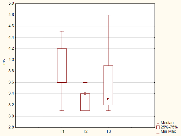
Fig. 1. Terminal latency in the “tarsus” stimulation point on the right with n. Peroneusstimulation in female martial artists before (T1), after the 1st session (T2) and after the magnetic stimulation course (T3)
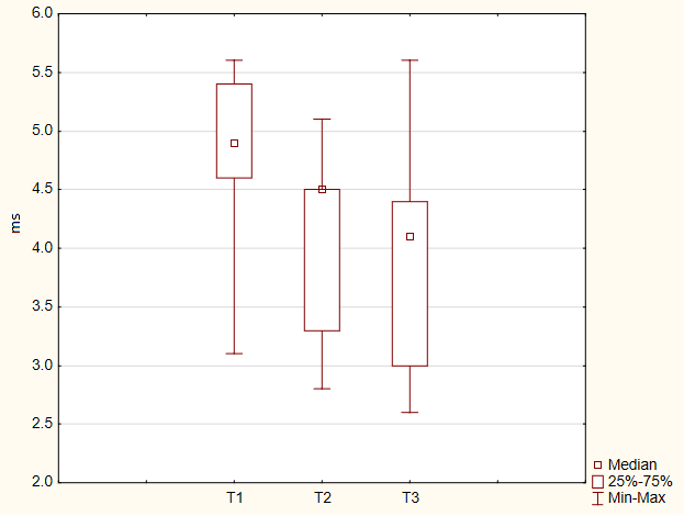
Fig. 2. Terminal latency in the “medial malleolus” stimulation point on the right with n. Tibialis stimulation in female martial artists before (T1), after the 1st session (T2) and after the magnetic stimulation course (T3)
After the magnetic stimulation course with n. Peroneus stimulation, the nerve conduction velocity decreased in the “popliteal fossa”stimulation point on the right (before – 56 (55.3;57.1) m/s, after the course – 50 (49.5;53.5) m/s, p<0.04) and on the left (before – 57.7 (56.8;60) m/s, after the course – 51.7 (49.5;56.2) m/s, p<0.04).
After the 1st magnetic stimulation session with n. Peroneus stimulation, the M-response area reduced in the “tarsus” point on the right (before – 23.65 (13.9;32.55) mV×ms, after – 16.6 (9.6;23.8) mV×ms, p<0.05) (fig. 3), in the “head of fibula” point on the right (before – 22.85 (14;31.05) mV×ms, after – 16.45 (8.8;25.15) mV×ms, p<0.05) and in the “popliteal fossa” point on the right (before – 23.4 (14.8;36.95) mV×ms, after – 19.25 (9.9;29.15) mV×ms, p<0.05) in female cyclic sports athletes.
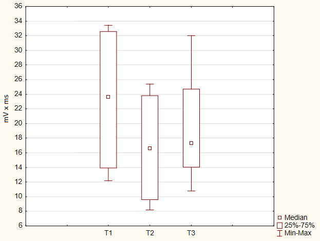
Fig. 3. M-response in the “tarsus” stimulation point on the right with n. Peroneus stimulation in female cyclic sports athletes before (T1), after the 1st session (T2) and after themagnetic stimulation course (T3)
After the magnetic stimulation course with n. Peroneus stimulation, the terminal latency decreased in the “head of fibula” point on the right (before – 10.7 (10.2;11.8) ms, after – 10 (9.75;10.55) ms, p<0.05). In the “popliteal fossa” point on the right, the M-response duration also decreased (before – 8.265 (6.89;8.975) ms, after – 7.85 (6.6;8.55) ms, p<0.05). When the n. Tibialis was stimulated, the M-response duration decreased as well in the “medial malleolus” point on the right (before – 6.46 (5.725;7.36) ms, after – 5.41 (4.9;6.01) ms, p<0.05).
Also, after the magnetic stimulation course with n. Peroneus stimulation, the terminal (before – 4 (3.5;4.7) ms, after – 3.6 (3.25;3.95) ms, p<0.05) and residual latency (before – 2.345 (1.935;2.645) ms, after – 2,345 (1.935;2.645) ms, p<0.05) decreased in the “tarsus” stimulation point on the left.
Analysis of changes in the peripheral blood flow of the lower limbs of women engaged in martial arts before, after the 1st session and after the magnetic stimulation course has revealed an increased rheographic index in the “left foot” segment (before – 0.57 (0.55;0.69) c.u., after the 1st session – 0,92 (0.81;1.09) c.u., after the course – 1.17 (0.69;1.4) c.u., p<0.04). In the “right foot” segment, after the 1st session, there was a statistically significant increase in the time of the maximum systolic filling of the vessels (before – 0.122 (0.1;0.126) s, after – 0,158 (0.141;0.17) s, p<0.04) and elastic modulus (vascular tone indicator) (before – 10 (10;13) %, after – 15 (12;23) %, p<0.04). After the 1st session, the rheographic wave propagation time (before – 0.24 (0.24;0.27) s, after – 0.26 (0.25;0.28) s, p<0.04) and the rime of slow filling of the vessels (before – 0,092 (0.079;0.095) s, after – 0.109 (0.104;0.122) s, p<0.04) decreased in the “left tibia” segment. After the course, the venous outflow significantly increased in the “left tibia” segment (before – 13 (12;29) %, after – 25 (22;47) %, p<0.05).
In cyclic sports athletes only statistically significant changes in the increase of the time of slow filling of the vessels were revealed in the “right foot” segment after the 1st magnetic stimulation session (before – 0.089 (0.074;0.103) s, after – 0.117 (0.1;0.155) s, p<0.05).
Conclusion. Therefore, repeated peripheral magnetic stimulation on the lumbar and sacral spine has caused unidirectional changes of the neuromuscular system of the lower limbs: reduction of M-response latency and duration, although the M-response area was also reduced. Rheovasography of the lower limbs in the “tibia” and “foot” segments revealed a significant improvement of all hemodynamic parameters. Greater changes in the functional state and hemodynamics of the lower limbs were observed in martial artists. The revealed changes show that high-intensity repeated peripheral magnetic stimulation of the lumbar and sacral spine causes relaxation (reduced activity) of the neuromuscular system of the lower limbs and improvement of hemodynamics, which contributes to accelerated processes of urgent recovery after intense sports activity. The tested method of applying the high-intensity magnetic stimulation may be recommended for athletes to recover after an intensive training day during the preparatory period of sports training.
Conflict of interest. The authors declare no conflict of interest.
REFERENCES
- Ter-Akopov G.N. Medical and biological support of the highest achievement sports: status and topical issues (article one). Resort medicine, 2023, no. 2, pp. 139-146. (in Russ.)
- Pomytkin A.N., Lebedeva I.S., Tikhonov D.V., Kaleda V.G. Rhythmic transcranial magnetic stimulation in the treatment of resistant depression in schizophrenia. S. Korsakov Journal of Neurology and Psychiatry, 2021, vol. 121, no. 5-2, pp. 99-105. (in Russ.)
- Novikova E.S., Rozanov I.D. Low-dose radiotherapy for chronic pain syndrome in specific and nonspecific musculoskeletal system diseases. Medical alphabet, 2019, vol. 1, no. 2, pp. 58-58. (in Russ.)
- Pushkar’ D.Yu., Kulikov A.G., Kasyan G.R., Kupriyanov Yu.A., Romikh V.V., Zakharchenko A.V., Voronina D.D., Yarustovskaya O.V., Zajtseva T.N. Extracorporeal magnetic stimulation of the pelvic floor neuromuscular system in urological practice. Russian Journal of the Physical Therapy, Balneotherapy and Rehabilitation, 2019, vol. 18, no. 4, pp. 264-276. (in Russ.)
- Kasumova A.A., Krasnikova V.V., Pospelova M.L., Fionik O.V., Alekseeva T.M., Samochernykh K.A. Possibilities of using the transcranial magnetic stimulation method in the treatment of chronic pain syndromes. Modern problems of science and education, 2021, no. 2, pp. 195-195. (in Russ.)
- Kutashov V.A., Ulyanova O.V. Application of transcranal magnetic stimulation within complex treatment of patients with ischemic stroke in the late recovery period from medical rehabilitation positions. Herald of physiotherapy and health
resort therapy, 2018, vol. 24, no. 3, pp. 73-80. (in Russ.) - Koryagina Yu.V., Abutalimova S.M., Kushnareva Yu.V., Nopin S.V., Mitin I.N. Peripheral magnetic stimulation of the torso musclesof athletes during intensive training activity.Modern Issues of Biomedicine, 2024, vol. 8, no. 3. DOI: 10.24412/2588-0500-2024_08_03_28. (in Russ.)
- Astafyeva D.S., Vlasov Ya.V., Strelnik A.I., Chigareva O.V., Markina E.A., Shishkovskaya T.I., Smirnova D.A., Gayduk A.Ya. Repetitive transcranial magnetic stimulation use in neuropathic pain with comorbid depression: a review of efficient
treatment protocols’ parameters. Neuromuscular Diseases, 2023, vol. 13, no. 2, pp. 20-30. (in Russ.) - Bakulin I.S., Pojdasheva A.G., Lagoda D.Yu., Evdokimov K.M., Sabirova A.Kh., Suponeva N.A., Piradov M.A. Safety and tolerability of different protocols of high-frequency rhythmic transcranial magnetic stimulation. Ulyanovsk Medico-biological Journal, 2019, no. 1, pp. 26-37. (in Russ.)
INFORMATION ABOUT THE AUTHORS:
Yulia V. Koryagina – Doctor of Biological Sciences, Professor, Head of the Center of Biomedical Technologies, FSBI “North-Caucasian Federal Research-Clinical Center of Federal Medical and Biological Agency”, Essentuki, e-mail:
Sergej V. Nopin – Candidate of Technical Sciences, Lead Researcher of the Center of Biomedical Technologies, FSBI “North-Caucasian Federal Research-Clinical Center of Federal Medical and Biological Agency”, Essentuki, e-mail:
Yulia V. Kushnareva – Candidate of Biological Sciences, Senior Researcher of the Center of Biomedical Technologies, FSBI “North-Caucasian Federal Research-Clinical Center of Federal Medical and Biological Agency”, Essentuki, e-mail:
Sabina M. Abutalmova – Candidate of Medical Sciences, Senior Researcher of the Center of Biomedical Technologies, FSBI “North-Caucasian Federal Research-Clinical Center of Federal Medical and Biological Agency”, Essentuki, e-mail:
Igor’ N. Mitin – Candidate of Medical Sciences, Leading Researcher, FSBI “Federal Research-Clinical Center of Sports Medicine and Rehabilitation of Federal Medical and Biological Agency”, Moscow.
For citation: Koryagina Yu.V., Nopin S.V., Kushnareva Yu.V., Abutalimova S.M., Mitin I.N. Functional status of the lower limbs after peripheral magnetic stimulation of the lumbar and sacral spine of athletes of different sports. Russian Journal of Sports Science: Medicine, Physiology, Training, 2024, vol. 3, no. 3(11). DOI: 10.24412/2782-6570-2024_03_03_6